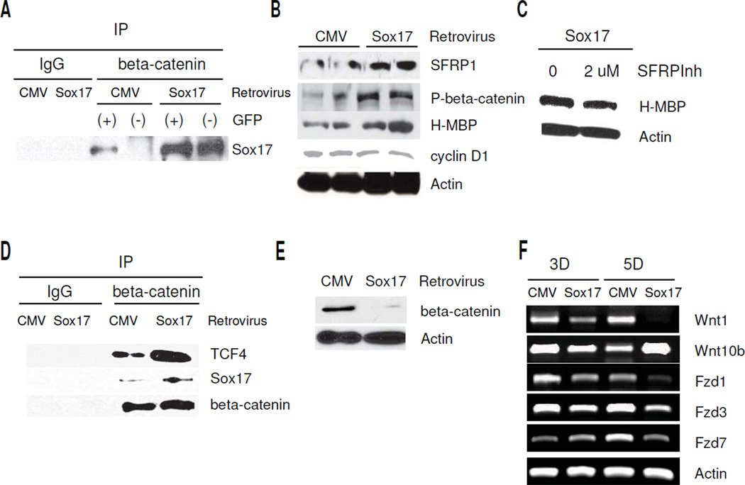
Figure 5.
Recombinant Sox17 induces beta-catenin-TCF complex formation in HOG cells, and decreases Wnt/beta-catenin signaling by cell autonomous and non cell-autonomous mechanisms. A. Sox17 retrovirus transduction increases Sox17 levels in beta-catenin immunoprecipitates of both GFP+ and GFP− cell populations. GFP+ and GFP− cells were isolated from CMV vector or Sox17-transduced HOG cells by FACS sorting 5 days after transduction. Lysates were prepared from the purified cells and immunoprecipitated with mouse IgG (CMV lysate only) or beta-catenin antibody. Western blotting was performed for Sox17. Sox17 levels are approximately 2.5 fold higher in Sox17 GFP(+) compared with CMV GFP(+). Note increased Sox17-beta-catenin complex formation in GFP- cells after transduction with Sox17 retrovirus, indicating non-cell autonomous activity. B. Western blot showing changes in SFRP1, H-MBP and phospho-S33/37/T41-beta-catenin (P-beta-catenin) at 3 days post-transduction with control (CMV) or Sox17 retrovirus. Two samples for each group are shown. C. Western blot showing inclusion of 2 uM SFRP1 inhibitor (SFRPInh) decreases the Sox17-induced H-MBP 5 days following retroviral transduction. D. Western blot showing increased Sox17 and TCF4 are detected in beta-catenin immunoprecipitates (IP) 5 days after HOG cell transduction with Sox17 retrovirus. Negative controls consisted of CMV-transduced cell lysate precipitated with IgG. E. Western blot showing the levels of total beta-catenin is clearly reduced at 5 days after retroviral transduction. F. Semi-quantitative RT- PCR results showing decreased Wnt1, Fzd1, −3, and −7 expression in HOG cells after transduction with the Sox17 retrovirus. Samples were analyzed after 3 days (3D) and 5 days (5D) recovery from virus addition.

