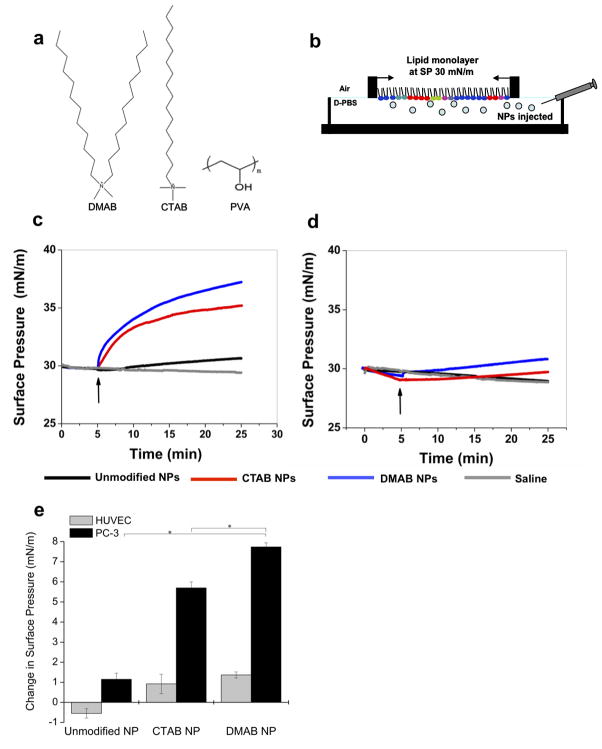
Figure 1. NP-Lipid biophysical interaction studies.
The different surfactants used in PLGA NP formulation (a). Unmodified NPs were formulated with PVA alone; surface-modified NPs were formulated with PVA and either DMAB or CTAB. To study NP-lipid interactions, representative lipid monolayers were created over a buffer subphase into which NPs were injected (b). The change in surface pressure (ΔSP) was monitored over time for PC-3 lipids (c) and HUVEC lipids (d). Curves represent the median result. The positive ΔSP at 20 min after NP injection demonstrates quantitatively that DMAB-NPs had greater interaction with PC-3 lipids than did CTAB- and unmodified NPs and that DMAB-NPs also had greater interaction with PC-3 cells than with HUVECs (e). Data are shown as mean ± standard deviation; n = 3;*p< 0.05.

