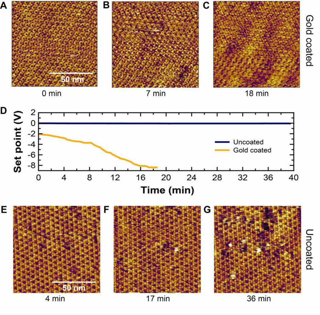
Fig. 7.
Time-lapse imaging of a 100 × 100 nm area (~4 Å/pixel) of a BR patch using a gold coated and uncoated BioLever Mini. (A–C) Images acquired at times 0, 7 and 18 min using a gold-coated BioLever Mini. (D) Plot showing the set point during imaging using gold-coated (gold) and uncoated (blue) BioLever Minis, respectively. (E–G) Images acquired at times 4, 17, and 36 min using an uncoated BioLever Mini.

