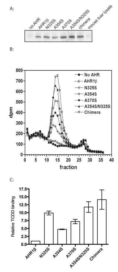
Figure 4. Mutation of specific LBD residues increases TCDD binding of AHR1β.
(A) Expression of each AHR in TNT reactions determined by western blotting. (B) Velocity sedimentation analysis of TCDD binding. Synthetic AHR proteins or unprogrammed TNT lysates were incubated with 2 nM [3H]TCDD and fractionated on sucrose density gradients. Sedimentation marker [14C]catalase (11.3S) eluted in fractions 14-20 of all gradients. The experiment was replicated three times; a single example is shown. (C) Quantification of TCDD specific binding revealed by sedimentation analysis. Radioactivity (dpm) in fractions comprising each peak in (B) was summed. Specific binding is the difference between total binding (preparations containing an AHR) and non-specific binding (preparation lacking AHR). Bar graph plots specific binding relative to that found for AHR1β. Values represent the mean ± standard error for 3 replicates.

