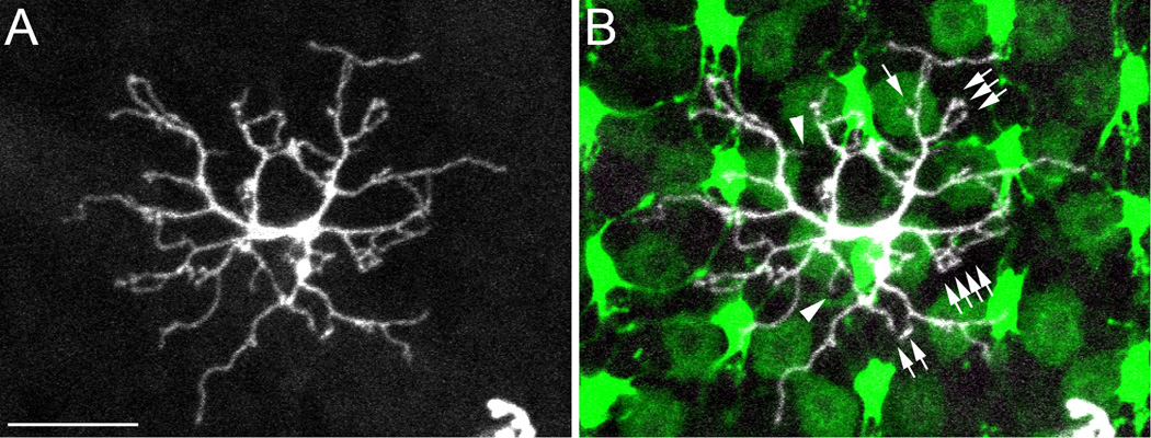
Figure 2.

A type specimen RGBUVRod bipolar cell in a SWS1-GFP:LWS-GFP double transgenic zebrafish retina. A: The z-axis projection of serial confocal images of a RGBUVRod bipolar cell showing its dendritic tree. B: Overlaying A with a single-plane confocal image of the photoreceptor terminals. UV and R cone terminals have bright and dim fluorescence, respectively. This bipolar cell connects to cone terminals with single, double, triple or more dendritic terminals (arrows). The dendritic terminals fall on R, G, B, and UV cone terminals except two that presumably connect to rod terminals (arrowheads). Scale bar = 10 µm.
