
Fig. 5.
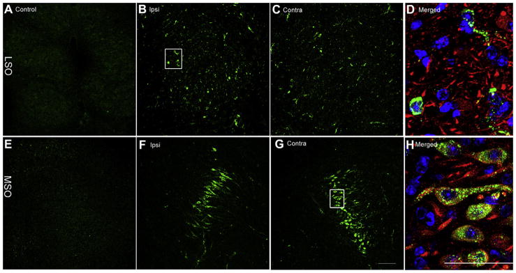
Bilateral distribution of gentamicin immunolabeling in the MSO and LSO 7 days following gentamicin administration. Negligible gentamicin immunolabeling is present in LSO (A) and MSO (E) from the saline control group. Scattered positive gentamicin immunloabeling was observed in the ipsilateral (B) and contralateral (C) LSO, with a focused band of labeled neurons in the ipsilateral (F) and contralateral (G) MSO. (D) and (H) Triple-labeled high-resolution images (of the boxed area in B and G, respectively) revealed that gentamicin immunolabeling (green) is mainly distributed within the cytoplasm and axons of neurons. Blue, DAPI; red, beta-III tubulin immunolabeling. Scale bar = 50 μm in A, B, C, E, F, and G, and 100 μm in D and H. (For interpretation of the references to color in this figure legend, the reader is referred to the web version of this article.)