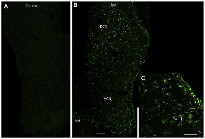
Fig. 6.
Gentamicin immunolabeling in the ipsilateral cochlear nucleus at 1 day after transtympanic administration. (A) Gentamicin immunolabeling is absent in the contralateral cochlear nucleus (Contra/contralateral, DCN/dorsal cochlear nucleus). This figure is a composite of images from one brain section spliced together. (B) Gentamicin immunolabeling (green) was extensive in the ipsilateral DCN (Ipsi/ispilateral) and ventral cochlear nucleus (VCN). The intensity of anti-GT immunostaining in the DCN is stronger than that in the VCN. This figure is a composite of images from one brain section spliced together. (C) Nerve fibers within the DCN also displayed gentamicin immunolabeling (white arrows). Images are from the same tissue sample, different section. Scale bar = 100 μm. (For interpretation of the references to color in this figure legend, the reader is referred to the web version of this article.)

