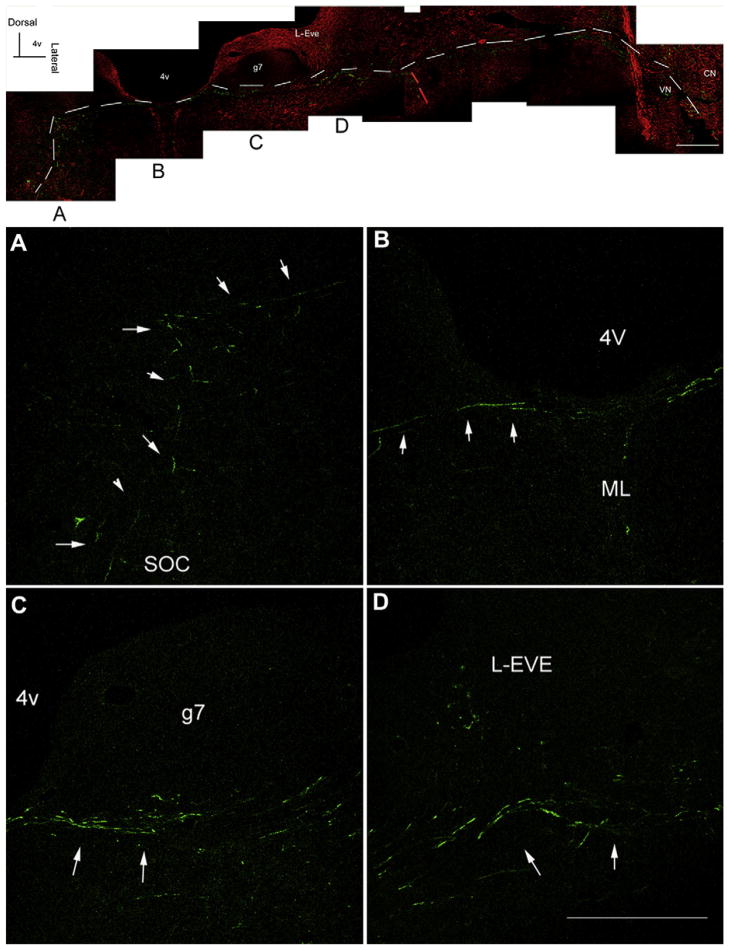
Fig. 9.
Gentamicin immunolabeling along the descending efferent pathways from the SOC 7 days after transtympanic administration. Adjoining images taken from the same section were spliced together to demonstrate the route of nerve fibers with gentamicin immunolabeling. White dashed lines indicate the route of gentamicin immunolabeled nerve fibers (green) following the typical route of the descending efferent pathways from the contralateral SOC, along the surface of the floor of fourth ventricle, to the root of the vestibular nerve. Red dashed lines indicate the route of gentamicin immunolabeled nerve fibers (green) along the descending pathway from the ipsilateral SOC to the root of vestibular nerve. Each lower panel magnifies the corresponding image in the respective upper panel. Red, beta-III tubulin immunolabeling. Scale bar is 300 μm. (For interpretation of the references to color in this figure legend, the reader is referred to the web version of this article.)

