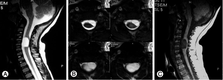
Fig. 1.
(A) T2 magnetic resonance (MR) images of case one; sagittal view showing hyperintense mass anterolateral to the cord at C5-C6 level. (B) Axial view, showing the left anterolateral location of the cyst. (C) T1-weighted MR image demonstrating hypointense cyst at C5-C6 level of the same patient.

