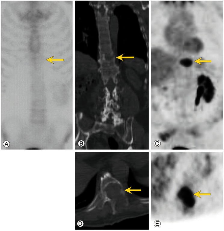
Fig. 2.
Bone scan (A), computed tomography (CT) (B, D) and fluorodeoxyglucose (FDG) positron emission tomography (PET) (C, E) images of a 69-year-old man with renal cell carcinoma. Note the focal intense uptake in osteolytic spine metastases (vertebral body of T10) on PET only but not on the bone scan (arrows). Axial and coronal CT images show osteolytic changes in the vertebral body (B, D) and focal intense uptake on the PET images (C, E).

