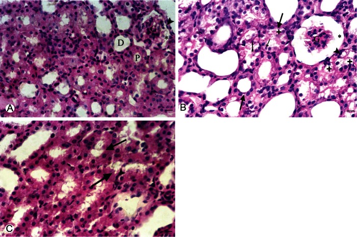
Figure 3.
A. Representive photomicroscopy of a section in the kidney of a control rat showing the renal corpuscle with the parietal layer of Bowmman’s capsule (arrow head). glomerular and normal Bowmann’s space (*). Normal appearance of the proximal (P) and distal (D) in control convulated tubules. B. Group II showing apparent glomerular atrophy (G) with widened Bowman’s space (*). Swelling of the renal tubules is observed (thick arrows). The proximal tubules showed marked vaculation of the cytoplasm (+), some nuclei in the proximal tubules appear pyknotic (arrow head) or karyolsed (thin rrow) and others show exfoliation (tailed arrow). C. Group III showing swelling of the renal tubules (thick arrows) and some proximal tubules showed pyknotic nuclei (arrow head). (H&E) X 400. Bar = 50 μm.

