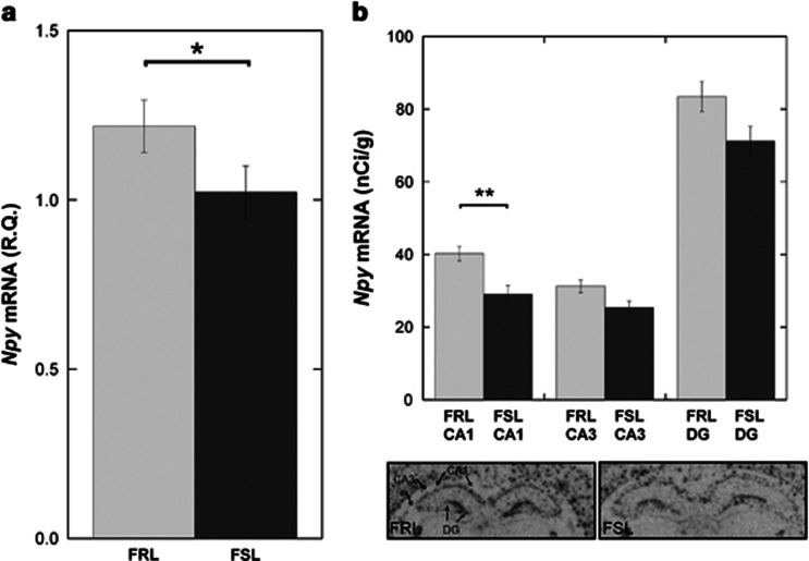
Figure 2.
Gene expression analyses of Npy in (a) whole hippocampal homogenate using quantitative real-time PCR (qRT-PCR) and (b) different hippocampal regions using in situ hybridization (representative in situ autoradiograms are shown below the figure). Analyses show a decrease in neuropeptide Y (Npy) mRNA levels, in the hippocampus of the Flinders sensitive line (FSL), which is most pronounced in the cornu ammonis (CA)1 area. *P<0.05, **P<0.01.

