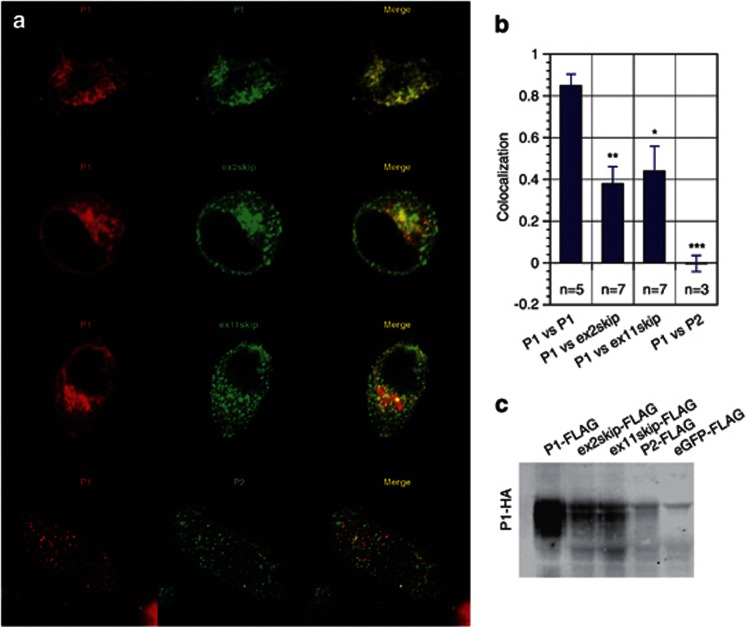
Figure 4.
EAAC1 isoforms partially colocalize and interact with the EAAC1 P1 protein in transfected HeLa cells. (a) HeLa cells were transfected with equal amounts of DNA corresponding to P1-HA-tag and to FLAG-tagged isoform (P1, ex2skip, ex11skip, or P2) as indicated. HeLa cells were used in this experiment because they do not express SLC1A1/EAAC1. The hemagglutinin (HA)-tag and FLAG-tag were detected using Alexa555-conjugated anti-rabbit and Alexa647-conjugated anti-mouse antibodies, respectively, and visualized using confocal microscopy as described in Material and methods. Confocal images were optimized for each channel (red=HA; green=FLAG) as indicated. (b) Quantitation (±s.e.m.) of colocalization for FLAG-tag and HA-tag for each of the combinations shown in a, using the method of Costes.36 *P⩽0.05, **P⩽0.01 and ***P⩽0.001 in comparison to the P1 vs P1 colocalization. (c) HeLa cells transfected with DNA corresponding to P1-HA-tag and the indicated FLAG-tagged isoform were lysed 2 days post transfection. Lysates were then bound to FLAG-beads, pulled down and proteins resolved by SDS-polyacrylamide gel electrophoresis. HA-tagged P1 was detected using anti-rabbit antibody (see Materials and methods).

