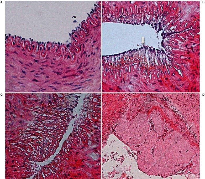
Figure 3.
Histopathological findings after mechanical thrombectomy. A) Microscopic view of a right SFA (control sample) shows all layers preserved (hematoxylin-eosin [H&E] staining, original magnification ×400). B,C) Microscopic view of an artery treated with the aspiration device demonstrate the intact internal elastic lamina (thin arrow) and a single layer of endothelial cells (thick arrow). The parentheses show edema of the subendothelial layer in B and the media layer in C. There is no thrombus (H&E staining, original magnification ×400). D) Microscopic view of an artery treated with the Catch thromboembolectomy system shows a mural clot (parenthesis) and the internal elastic lamina fractured. (H&E staining, original magnification ×200).

