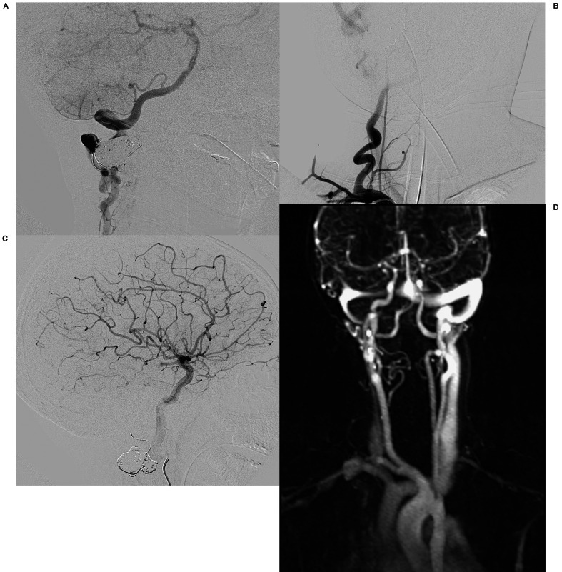
Figure 4.
A-C) Follow-up angiography 6 weeks after embolization. A) Right deep cervical artery angiography lateral view demonstrates a decrease in size of the previously enlarged deep cervical artery with reconstitution of the right vertebral artery at the C2 level. Collateral branches fill the distal V3 and V4 segments of the right vertebral artery in antegrade fashion and there is antegrade flow within the basilar artery with a normal posterior circulation. B) Right subclavian artery AP angiogram demonstrates opacification of the diminutive proximal cervical vertebral artery extending just proximal to the level of the treated arteriovenous fistula. There is no evidence for any supply to the arteriovenous fistula. C) Lateral view of the right internal carotid artery angiography reveals normal distal cervical and intracranial segments of the artery without opacification of the basilar artery consistent with antegrade flow within the basilar artery. D) Follow-up time-resolved MR angiography 15 months after embolization demonstrates no early venous enhancement consistent with the resolution of the arteriovenous fistula.

