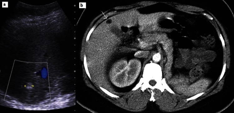
Figure 3.

(A) Ultrasound of the liver showed well-circumscribed hyperechoic lesions (callipers). (B) which were of fat density (≤10 Hounsfield units; arrow) on CT, consistent with angiomyolipomas. Surgical absence of left kidney noted (owing to previous ruptured angiomyolipoma). Normal right kidney.
