
Figure 1.
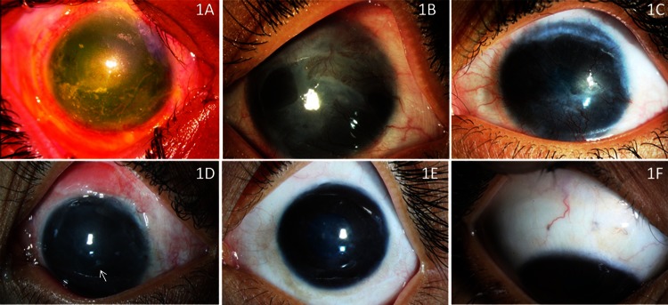
(A) Clinical photograph of the left eye showing extensive corneal limbal and conjunctival epithelial defect with corneal stromal haze, 3 days following lime injury. (B) Same eye 6 months after amniotic membrane grafting, showing conjunctivalised ocular surface, suggestive of limbal stem cell deficiency (LSCD). (C) Same eye 1 year after autologous cultivated limbal epithelial transplantation (CLET), showing a vascular and conjunctivalised ocular surface suggestive of recurrence of LSCD following CLET. (D) Postoperative photograph of the left eye 3 weeks after autologous simple limbal epithelial transplantation (SLET), showing an epithelised corneal surface with few limbal transplants in place (arrow). (E) One year post-SLET the left eye shows a stable, epithelised and avascular corneal surface with significant improvement in corneal clarity. The corrected visual acuity at this stage was 20/80. (F) Right eye showing healthy donor sites with no ocular surface deficits.