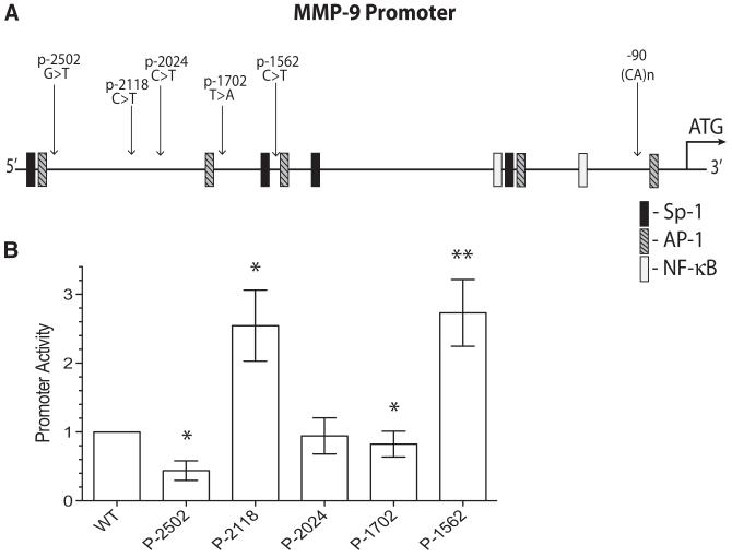
Figure 2.
Basal promoter activity of MMP-9 promoter SNPs. A, A cartoon of the MMP-9 genomic sequence upstream of the start methionine depicting the relative location of the 5 promoter SNPs and the (CA)n microsatellite polymorphism. Locations of the DNA consensus binding motifs for some of the common transcription factors are also noted. B, Relative promoter activity normalized to the wt promoter–reporter construct. HEK293 cells transfected with the noted promoter–reporter constructs in a 24-well dish (0.4 μg promoter–reporter plasmid + 0.013 μg Renilla plasmid/well) were harvested 24 hours later and replated into 3-replicate wells of a 96-well plate at an approximate cell density of 104 cells/well in DMEM supplemented with 1% FBS. The firefly luciferase promoter–reporter activity normalized by the Renilla luciferase activity to compensate for potential transfection efficiency and cell density differences were assessed 24 hours later. Bars represent mean±SEM from 6 independent experiments each with triplicate readings. *P<0.05 and **P<0.01 by Mann–Whitney nonparametric test. MMP-9 indicates matrix metalloproteinase-9; SNP, single nucleotide polymorphism; HEK, human embryonic kidney; FBS, fetal bovine serum.

