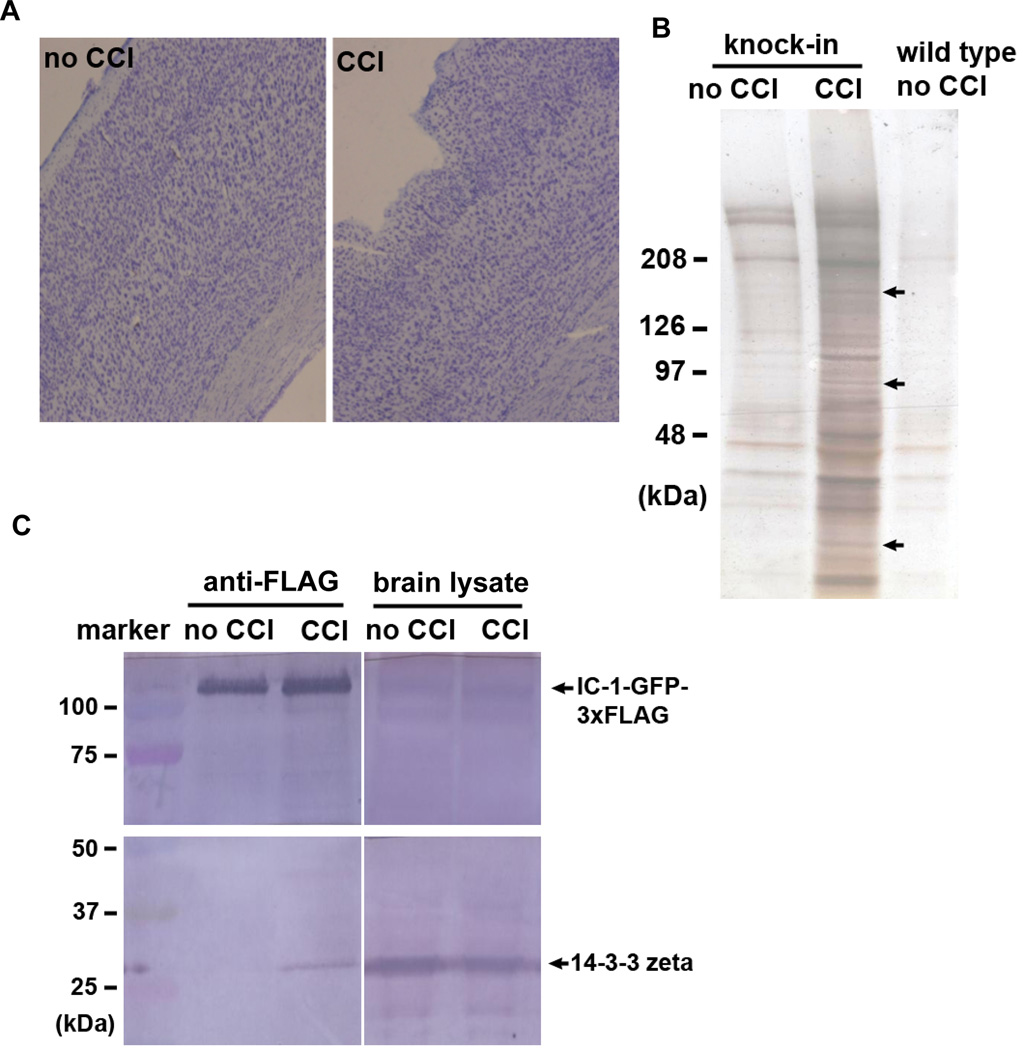
Figure 5.
Isolation and identification of proteins that were pulled down with IC-1-GFP-3xFLAG after brain injury. (A) Images of brain sections stained by cresyl violet showing the injury site of the mouse brain cortex after the Controlled-Cortical-Impact (CCI) procedure. (B) A silver-stained gel showing bands of pulled-down proteins from the injured brain. Three of these bands (indicated by arrows) were analyzed by mass spectrometry. Data of the two top hits from each band are shown in Table 1. (C) A western analysis on brain lysate and proteins pulled down by anti-FLAG antibody. The western blot was probed with the anti-14-3-3 zeta/delta antibody.

