
Fig. 1.
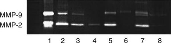
Gel zymogram depicting differences in (pro-)MMP-2 and (pro-)MMP-9 content among discrete samples of glial cells, serum supplements, and cerebral tissue. After activation, the gel was stained with Coomassie blue by the methods outlined here (note that a parallel gel stained with amido black was more sensitive with regard to the samples with larger contents of gelatinase, suggesting a different slope of linearity of that system.). The samples in the individual lanes are: Lane 1, human recombinant MMP-2 and MMP-9. Lane 2, fetal bovine serum (FBS, 2%). Lane 3, serum-free medium in the well that had previously been incubated with 10% FBS-containing medium. Lane 4, astrocytes (from C57 Bl/6 mice) followed by PBS wash prior to serum-free medium; note the single pro-MMP-2 band. Lane 5, astrocytes without PBS wash prior to serum-free medium; note the presence of both a pro-MMP-9 band (bovine) and a pro-MMP-2 band (murine). Lane 6, microglia (from C57 Bl/6 mice) followed by PBS wash prior to serum-free medium. Note the single pro-MMP-9 band. Lane 7, microglia without a PBS wash prior to serum-free medium. Note that the MMP-9 “doublet” represents pro-MMP-9 from both murine (cell) and bovine (FBS) sources. The lower band of the doublet is not active murine MMP-9, but derived from FBS. Lane 8, sample of cortical tissue (Papio anubis cynocephalus) taken 7 days after middle cerebral artery occlusion. Note the faint bands of pro-MMP-2 and pro-MMP-9.