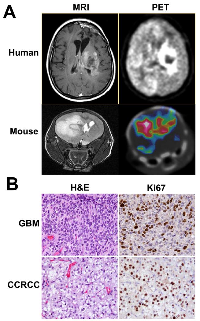
FIGURE 2.
Orthotopic primary GBM mouse tumors share MR and PET imaging features seen in original patient. In panel A, both human and orthotopic mouse GBM tumors appear as a large hyperintense T2 mass, with significant mass effect (mouse MRI). Clinical and orthotopic tumor FDG-PET images are consistent with enhanced glucose uptake relative to the surrounding brain tissue. Panel B shows hematoxylin-eosin (H&E) and Ki67 staining from the orthotopic GBM and orthotopic CCRCC brain metastasis. GBM tumors were characterized by a high mitotic index, pleomorphic nuclei, tortuous microvasculature and diffuse single cell infiltration. In contrast, the CCRCC showed brisk proliferation, monomorphic nuclei and scant cytoplasm. Magnification of histological images is 20×.

