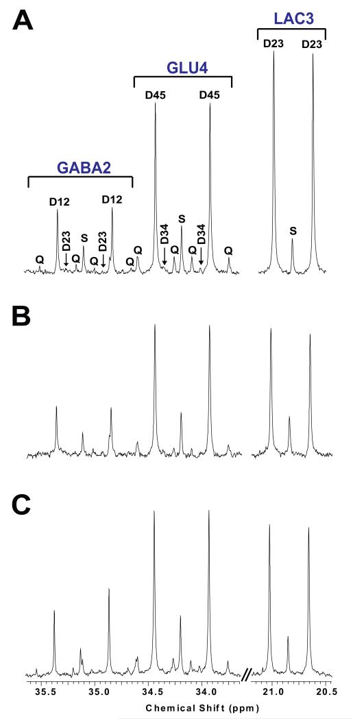
FIGURE 4.
Section of the 13C-NMR spectrum illustrating the labeling pattern of glutamate C4 (GLU4), GABA C2 (GABA2), and Lactate C3 (LAC33) in normal forebrain (A), surrounding brain of GBM (B), surrounding brain of CCRCC metastatic to the brain (C). The labeling pattern between both isotopomers is notably consistent throughout all brain tissues. S: singlet, Dxx: doublet, Q: quartet.

