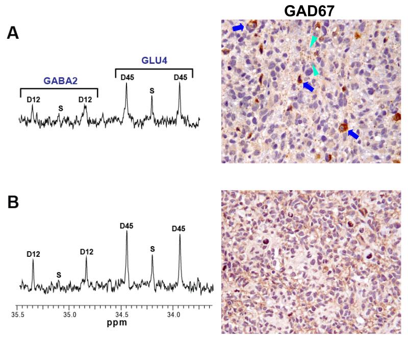
FIGURE 5.
Section of the 13C-NMR spectrum showing the labeling pattern of glutamate C4 (GLU4) and GABA C2 (GABA2) in GBM (A) and CCRCC orthotopic tumors (B) and their respective immunohistochemical staining for GAD 67 (20×). In contrast to GBM, in the CCRCC orthotopic tumor GABA2 showed a lower singlet-to-doublet ratio relative to its precursor GLU4, consistent with compartmentalization of GABA synthesis in the tumor mass. In addition, CCRCC was homogeneously immunopositive for GAD67 throughout the tumor whereas GBM cells were immunonegative, suggesting that CCRCC cells are able to produce GABA, and that the GABA2 observed in the GBM 13C spectrum is derived from GABAergic interneurons (blue arrows) and GABAergic projections (green arrowheads) interspersed within the tumor tissue.

