
Figure 1.
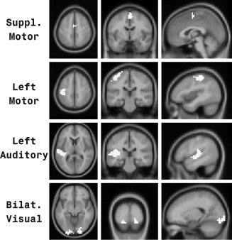
ROIs. Four of six ROIs employed in this study. ROIs are shown in white. For each ROI, an axial, coronal, and sagittal view is presented. The remaining two ROIs (right motor and right auditory) were comparable to the contralateral ROIs shown in this figure.