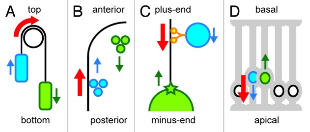
Figure 1. A funicular and cellular funiculars. The directions of active force generation are indicated in red. Blue and green indicate each of the two components moving in opposite directions (the directions are shown in arrows). The blue components are more directly linked to the active force generators, while the green components are moved more passively. (A) A funicular. Blue and green vehicles are linked by a cable. By applying a force to rotate the pulley at the top, the blue vehicle moves up, and the green vehicle moves down. (B) Cytoplasmic streaming in C. elegans. The molecular motor myosin generates forces at the cell cortex (black line) to move the proteins and granules toward the anterior (cortical flow, blue). The hydrodynamic property of the cytoplasm transmits the force to the inner components to move them posteriorly (cytoplasmic flow, green). (C) Centrosome centration in C. elegans. Organelles (blue) move toward the minus-end of a microtubule (black line) driven by the molecular motor dynein (orange). These movements generate a reactionary force that pulls the microtubule and associated centrosome (green star) and nucleus (green circle) toward the plus-end. (D) Interkinetic nuclear movement during mouse neurogenesis. Inside the developing brain, neural progenitor cells (gray cells with processes at their apical and basal surfaces) are densely packed. The nucleus in a G2-phase cell (blue) moves toward the apical surface using the force generated by the microtubule motor dynein. As the apical side gets crowded with the nuclei, the nucleus in the G1-phase cell (green) is pushed out toward the basal side.
