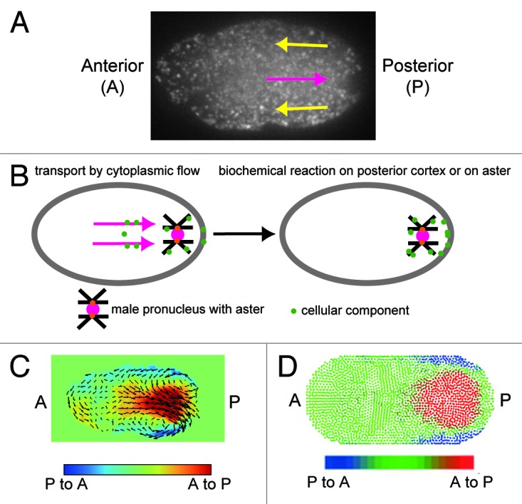
Figure 2. Cytoplasmic streaming in C. elegans. (A) Cytoplasmic streaming visualized using GFP-labeled yolk granules. Anterior-directed cortical flow and posterior-directed cytoplasmic flow are indicated by yellow and magenta arrows, respectively. (B) Our model for the possible role of cytoplasmic flow. The flow may enhance the probability of cytoplasmic material attaching to the aster or the posterior cortex. (C) Velocity distribution of cytoplasmic streaming was measured using the PIV method and visualized with vectors and colors.13 In the red region, the flow is posterior-directed, while in the blue region, the flow is anterior-directed. (D) Velocity distribution of streaming reproduced with computer simulation utilizing the MPS method.13 In the red region, the flow is posterior-directed, while in the blue region, the flow is anterior-directed.
