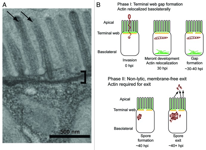
Figure 2. Intestinal cell morphology and microsporidia life cycle. (A) Electron micrograph of C. elegans intestinal epithelial cell. The microvilli brush border (arrows) lining the lumen is prominent on the apical surface of the cell. Microvilli are anchored to the cell with the terminal web (bracket), which is visible as a line below the microvilli. Both of these morphological features are conserved with human intestinal cells. (B) Phases of N. parisii exit strategy from C. elegans host intestinal cells. Green is ACT-5 and orange is IFB-2. Pathogen cells are depicted in red and nuclei depicted in black. During Phase I of infection, microsporidia spores fire their polar tubes, and inject their nucleus and sporoplasm into the host cell. This material develops into a multinucleated meront, and ACT-5 appears basolaterally, where it can form filament-like structures. During the final step of Phase I, gaps form in IFB-2 expression along the apical part of the cell. During Phase II, spores mature at approximately 40hpi and exit in a non-lytic, actin-dependent fashion from the host cell into the intestinal lumen.

An official website of the United States government
Here's how you know
Official websites use .gov
A
.gov website belongs to an official
government organization in the United States.
Secure .gov websites use HTTPS
A lock (
) or https:// means you've safely
connected to the .gov website. Share sensitive
information only on official, secure websites.
