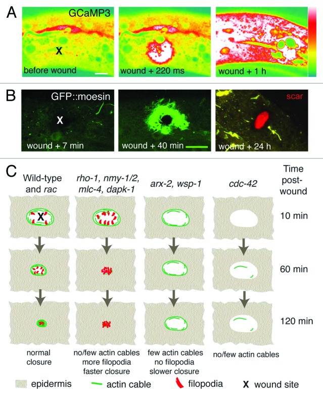
Figure 2.C. elegans epidermal wound responses. (A) Epidermal GCaMP fluorescence elevation after femtosecond laser wounding. Pcol-19-GCaMP3(juIs319). Lateral views of adult epidermis in mid-body before, immediately after and 1 h after laser wounding; x marks site of laser wound. Spinning disk confocal, intensity code; scale, 10 μm. (B) Needle wounding triggers actin assembly around the wound site. Pcol-19-GFP::Moesin (juIs352) labels actin filaments in adult epidermis. At 24 h an autofluorescent “scar” (red) is visible at the wound site and the actin structures have disappeared. x marks site of needle wound; laser scanning confocal images; scale, 10 μm. C, Graphical summary of mutant or RNAi phenotypes of genes implicated in C. elegans wound-induced actin dynamics.

An official website of the United States government
Here's how you know
Official websites use .gov
A
.gov website belongs to an official
government organization in the United States.
Secure .gov websites use HTTPS
A lock (
) or https:// means you've safely
connected to the .gov website. Share sensitive
information only on official, secure websites.
