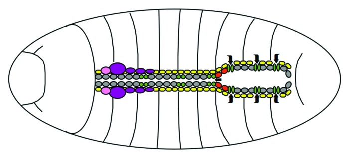
Figure 1. Schematic outline of the Drosophila heart. In the late stage embryo, cardioblasts (gray) shape the anterior tubular aorta and the posterior lumen of the dorsal vessel, interspersed with ostia (green) and aortic valves (red) to allow hemolymph flow (indicated by arrows). Adjacent pericardial cells (yellow) provide support and excretory functions. The lymph gland (purple) and ring gland (pink) lie associated to the cardiac system. Dorsal view, anterior is to the left.
