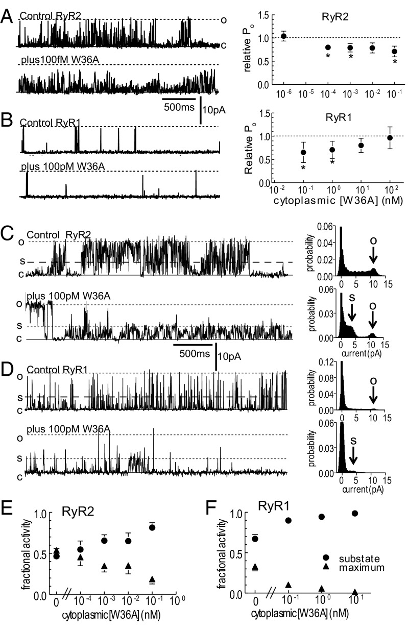
Fig. 3.
Activity of the mutant W36A toxin on RyR2 and RyR1 channels. (A and B) Cardiac RyR2 (A) and skeletal RyR1 (B); single channel currents at +40 mV (Left) and relative Po (Right), at indicated W36A concentrations. (C and D) Single RyR2 (C) and RyR1 (D) channel currents at +40 mV (Left) showing prolonged substate openings at indicated W36A concentrations. (Right) All points histograms. The arrow O indicates the full conductance level, and S points to the predominant substate current level. (E and F) The fraction of openings to submaximal levels (circles) and to the maximum conductance (triangles) for RyR2 (E) and RyR1 (F). Data are shown as mean ± 1 SEM. For visual clarity, only one error bar is shown. *Significant change at each toxin concentration.

