
Fig. 3.
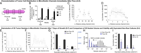
Characterization of tumor cell distribution in microfluidic channels immediately after flow (A–D), distribution of 3D tumor nodules in microfluidic channels after 7 d of growth (E and F), and comparison of 3D nodule volume and viability with equivalent nonflow cultures (G–I). (A) Intrachannel distribution of adherent ovarian cancer cells immediately after the introduction of three initial cell concentrations (104, 105, and 106 cells/mL) into the channels was quantified in three regions: proximal to the inlet (1.6–6.5 mm), middle (11.4–14.7 mm), and proximal to the outlet (19.4–24.3). (B) A concentration-dependent increase in the intrachannel tumor cell adherence density was observed in all three regions (P < 0.05). Within each initial cell concentration, results indicated no statistically significant difference in adherent cell densities across the channel in the three regions analyzed (P > 0.05). There was a trend toward decreased cell adherence density distal to the outlet at the initial cell concentration of 106 cells/mL. (C) Characterization of the number of adherent cells per linear mm [λ (x)] showed similar trends as a function of distance along the channel at the initial concentration of 106 cells/mL. (D) The number of cells adhered per linear distance across the channel [λ (y)] indicated higher concentration toward the channel center. (E and F) At an initial concentration of 106 cells/mL, adherent ovarian cancer cells that grew into 3D micronodules under the influence of continuous flow for 7 d were distributed along the full length of the channel (x axis) and across the width of the channel (y axis). (G) Compared with nonflow 3D cultures at all equivalent plating densities (black bars), growth under continuous flow (blue striped bars) resulted in a significant decrease in mean tumor volume. ***P < 0.001, *P = 0.01 to < 0.05. (H) A shift toward smaller tumors was observed in 3D nodules cultured under continuous flow (blue bars) compared with equivalent nonflow 3D cultures (white bars). (I) At all equivalent plating densities, 3D nodules grown under continuous flow (blue striped bars) had a significantly lower fractional viability than corresponding nonflow cultures (black bars). ***P < 0.001.