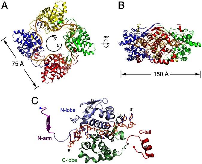
Fig. 3.
Structure of the BUNV NP–RNA complex. The tetrameric ring of the BUNV NP–RNA complex is shown as a ribbon diagram from the top (A) and side (B) views. Each protomer is presented as a colored cartoon, and the bound RNA is shown as a yellow cartoon. The bound RNA presents a 5′–3′ clockwise orientation. The molecular dimensions are also indicated. (C) The structure of the BUNV NP monomer. The N arm, N lobe, C lobe, and C tail are shown in magenta, blue, green, and red, respectively. The N and C termini are indicated. The bound RNA is shown as colored sticks.

