Fig. 5.
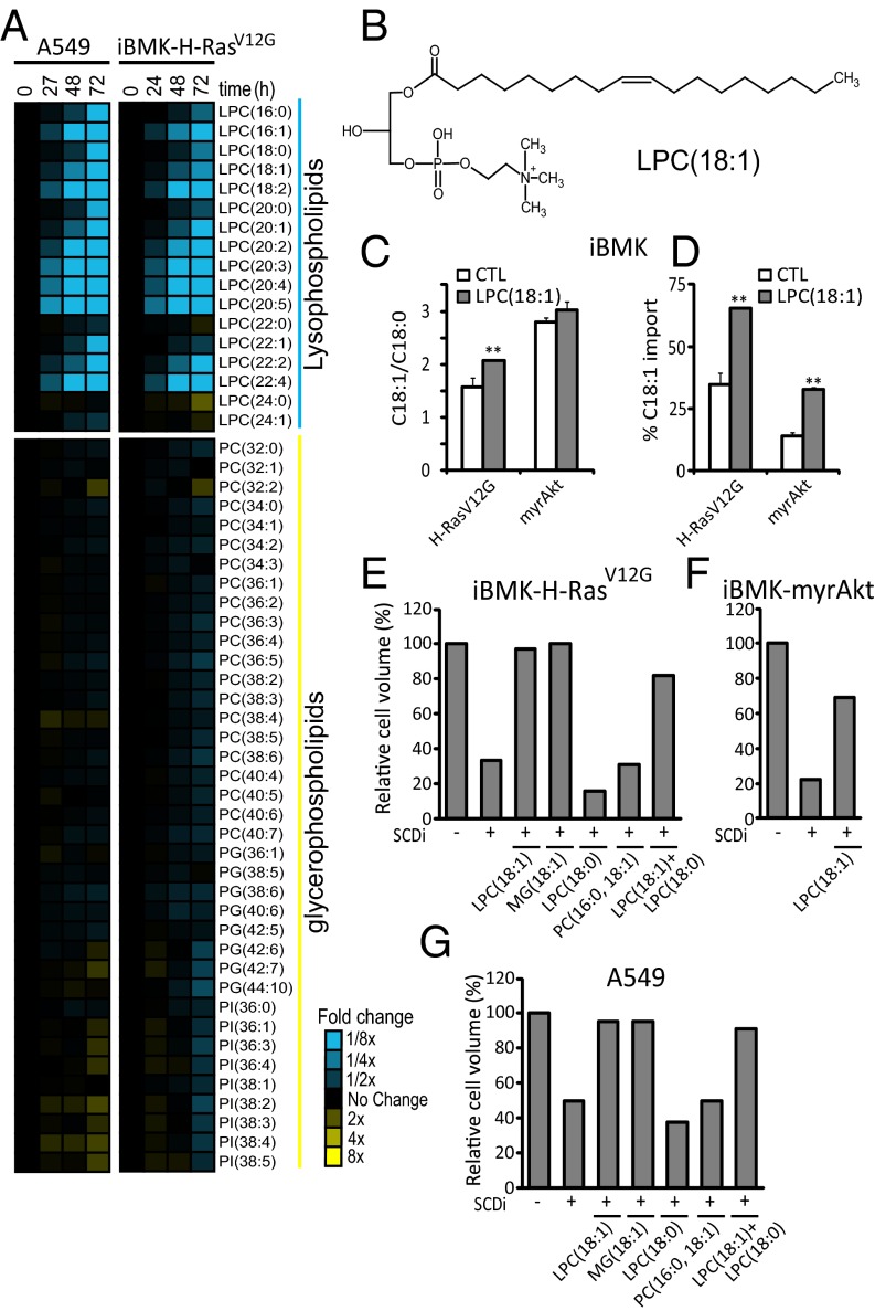
Fatty acids are scavenged from lysophospholipids. (A) Fold changes in medium phospholipids during growth of A549 and iBMK–H-RasV12G cells (relative to fresh medium with 10% serum). (B) Structure of LPC(18:1). (C) Effect of LPC(18:1) supplementation (20 µM; 72 h) on desaturation index (C18:1/C18:0). (D) Percentage of contribution of import to the cellular C18:1 pool based on [U-13C]glucose and [U-13C]glutamine labeling in iBMK–H-RasV12G and iBMK-myrAkt cells. (E–G) Packed cell volume for iBMK–H-RasV12G (E), iBMK-myrAkt (F), and A549 (G) cells [relative to untreated control (CTL)] after 72 h of incubation with or without 200 nM SCDi (CAY10566), in the presence of the indicated supplemented lipids (20 µM). PG, phosphatidylglycerol; PI, phosphatidylinositol. Data are mean n = 3 (A); means ± SD of n = 3 (C and D), and mean n = 2 (E–G). *P < 0.05; **P < 0.01 (two-tailed t test).

