Fig. 5.
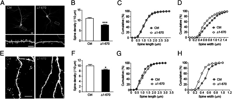
Disruption of CDKL5–PSD-95 interaction inhibits dendritic spine growth. (A) Representative images of hippocampal neurons transfected at DIV 8 with GFP together with empty vector or Flag-CDKL5-Δ1-670. Cells were fixed at DIV 15 and stained for GFP. [Scale bar: 20 μm (Top) and 5 μm (Bottom).] (B) Quantification of spine density in neurons transfected as indicated. n = 20–22 neurons for each; ***P < 0.001; t test. (C) Cumulative distribution of spine length plotted for the indicated conditions. P = 0.2586. Kolmogorov–Smirnov (K–S) test. (D) Cumulative distribution of spine width plotted for the indicated conditions. P < 0.001; K–S test. (E) Representative images of layer II–III pyramidal neurons in postnatal day (P) 14 rat brains transfected with GFP or GFP-CDKL5-Δ1-670 by in utero electroporation. Cells were stained with saturated GFP antibody to circumvent uneven distribution of GFP or GFP-tagged proteins in spines. (Scale bar: 10 μm.) (F) Quantification of spine density in neurons transfected as indicated. n = 5–6 neurons; *P < 0.05; t test. (G) Cumulative distribution of spine length plotted for the indicated conditions. P = 0.0414; K–S test. (H) Cumulative distribution of spine width plotted for the indicated conditions. P < 0.001; K–S test.

