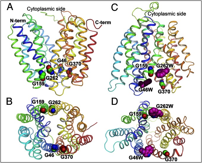
Fig. 1.
Trp replacements in two pairs of Gly–Gly residues that connect the N- and C-terminal six-helix domains on the periplasmic side of LacY. The 12 transmembrane helices that make up LacY are rainbow colored from blue to red. Gly residues (Gly46, 159, 262, and 370 in helices 2, 5, 8, and 11, respectively) and Trp replacements are shown as spheres. Inward-facing X-ray structure (Protein Data Bank ID code 2CFQ) viewed from the side (A) or from the periplasm (B). Model of the outward-facing conformation of LacY (18, 24) with Gly→Trp replacements (pink spheres) at positions 46 and 262 viewed from the side (C) or the periplasm (D).

