
Figure 2.
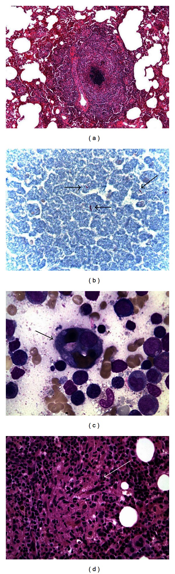
(a) Necrotizing granuloma evident on wedge-resected lung tissue. (b) Ziehl-Neelsen stain revealing acid-fast bacilli in resected lung tissue, consistent with mycobacteria (arrows). (c) Bone marrow aspirate specimen showing a hemophagocyte containing erythrocytes and pronormoblasts (arrow) (Wright stain, 1000x). (d) Core bone marrow showing a loose Granuloma (arrow) (Hematoxylin-Eosin stain, 400x).