
Figure 3.
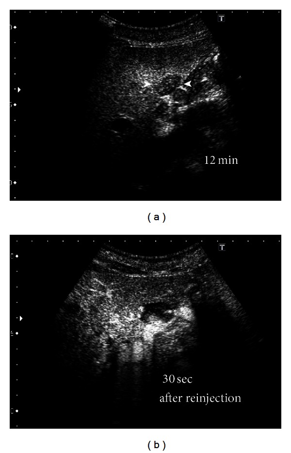
US, employing the “defect reperfusion US imaging” technique with Sonazoid, was performed on another hepatic hemangioma, in the left lobe of the liver, in tumor 1. (a) Preinjection image shows a hypoechoic mass measuring about 2 cm (arrowhead). (b) The lesion shows “peripheral-nodular enhancement” 30 s after reinjection of Sonazoid.