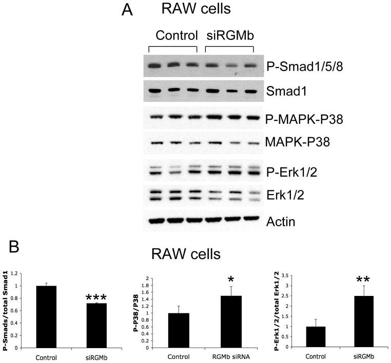
Figure 3.
Changes in phosphorylation levels of Smad1/5/8, and p38 and Erk1/2 MAPK in Dragon knockdown RAW264.7 cells. (A) RAW264.7 cells were transfected with control (three replicates) and Dragon siRNA (three replicates). 46 hrs after transfection, the cells were lysed and analyzed by Western blotting for phospho-Smad1/5/8 and total Smad1; phospho-p38 and total p38; phospho-Erk1/2 and total Erk1/2; and actin. (B) Western blot chemiluminescence from experiments in (A) was quantified using IPLab Spectrum software for phospho-Smad1/5/8 relative to Smad1, phospho-p38 relative to total p38, and phospho-Erk1/2 relative to total Erk1/2. *, p < 0.05; **, p < 0.01; ***, p < 0.001.

