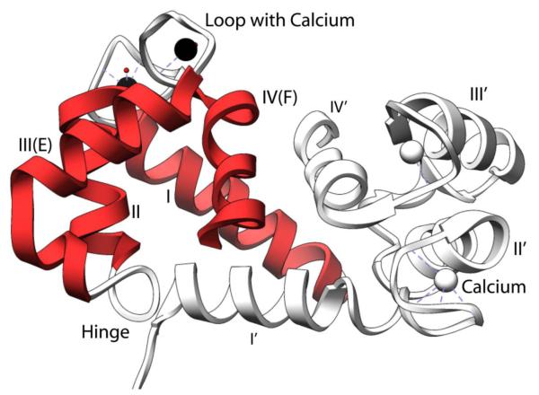
Figure 1.
Ribbon diagram of homodimeric calcium bound S100A9 from protein data bank file 1IRJ using the program Chimera.[113] Depicted in red is one subunit with the EF hand labeled. Secondary structure elements of homodimeric S100A9 include: helix one Q7-S23; helix two Q34-D44; hinge region L45-N55, calcium binding loop one: V24-N33, helix 3 E56-L66 (helix E of canonical EF Hand), calcium binding loop two D67-S75, and helix 4 F76-M94 (helix F of canonical EF hand). The C-terminal tail is disordered in the crystal structure and is therefore not shown in the figure. In this region, H92-E97 is the zinc binding motif. In the C-terminal disordered tail residues 103 to 105 represent the arachidonic acid binding region. There is also a truncated form of S100A9 of 12.7 kDa that is missing residues 1-4 found but has unclear biological function[114].

