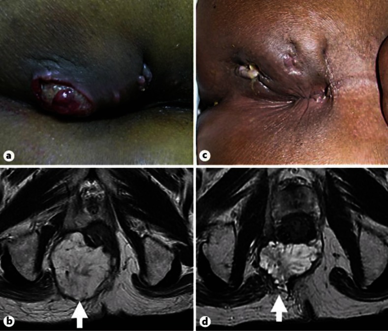
Fig. 1.
Time series presentation of physical examination and MRI. a, b Assessment before start of preoperative radiotherapy. Initial physical examination revealed an indurated, ulcerative lesion 7 cm in diameter with an external anal fistula opening (arrow). Initial MRI demonstrated a large demarcated tumor at the level of the anorectal junction with extension to the right side of the ischiorectal fossa and presacral space. c, d Assessment at the end of preoperative radiotherapy. The tumor had shrunk remarkably and the patient had a good clinical response based on imaging (arrow).

