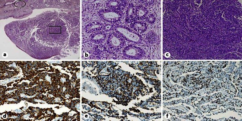
Fig. 1.
Specimen removed from the uterine cervix shows MANEC. a Intimate admixture of adenocarcinoma and NEC. HE. ×12.5. b Representative section of the tumor shows the adenocarcinoma component. A higher-magnification image of the oval area in a. HE. ×200. c Representative section of the tumor showing the NEC component. A higher-magnification image of the rectangular area in a. HE. ×100. d, e Tumor cells showing positive immunohistochemistry for synaptophysin, ×200 (d) and chromogranin A, ×200 (e). f MIB-1 proliferative index was approximately 25%. ×100.

