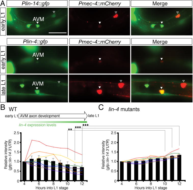
Fig. 7. Dynamic regulation of lin-14 3′UTR activity during AVM axon guidance in the L1 stage.

(A) Temporal expression of Plin-14::GFP and Plin-4::GFP reporters in the AVM neurons. An early L1-stage animal (top panel) showing lin-14 strongly expressed in AVM. An early L1-stage animal (middle panel) showing lin-4 weakly expressed in AVM and a late L1-stage animal (lower panel) showing lin-4 strongly expressed in AVM. The Pmec-4::mCherry reporter was used to label the AVM neurons. The merged images identify the GFP expressing cells as the AVM neurons. Arrowhead indicates the AVM neuron and the open arrowhead marks the ALM neuron. Anterior is to left, dorsal up. Scale bar, 20 μm. The expression intensity of the GFP::lin-14 3′UTR sensor in the AVM neurons was measured every hour at the first larval stage, starting at 4 hours after hatching and ending at 12 hours after hatching, in wild-type animals (B) and lin-4 mutants (C). Eight animals each were measured for wild type and lin-4 mutants. Bars represent the average intensity. ** and *** indicate intensity between wild type and lin-4 mutants is significantly different at P < 0.01 and P < 0.001, respectively. P values were calculated using a Student’s t-Test.
