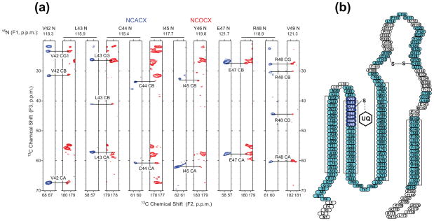
Figure 1.
SSNMR chemical shift assignments of DsbB (Cys41Ser). (a) Strip plot of NCACX (blue) and NCOCX (red) of DsbB (Cys41Ser) acquired at −12 °C, showing the residues at the active site of DsbB. (b) Topology view of DsbB with assigned residues highlighted in blue and cyan. Residues in blue are at the active site of DsbB. The four rectangles represent the transmembrane helices predicted by secondary structure analysis based on chemical shifts.

