
Fig. 1.
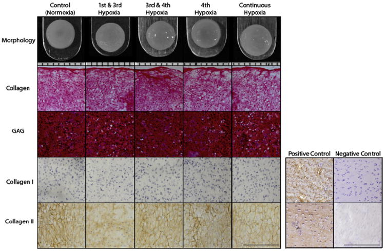
Gross morphology, histology, and IHC of self-assembled neocartilage. Both controls and hypoxia-treated constructs formed uniform neocartilage constructs with similar flat surfaces without physical abnormalities. The orientation of cryosectioning was from top to bottom in all groups (14 μm sections). Safranin-O/fast green staining for GAGs and Picrosirius red staining for collagen showed that constructs produced these matrix components uniformly for all groups. IHC illustrated that all groups were positive stained for collagen type II but not for collagen type I, suggesting AC phenotype. For gross morphology (top row), each notch represent 1 mm. For histology, bar represents 200 μm.