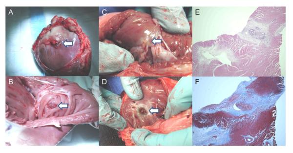
Figure 7. Right Ventricular Free Wall: Gross Pathology and Histopathology.

(A) Vascular closure device (collagen plug is visible; white arrow) deployed on epicardial surface at 2 days. (B) Vascular closure device (copolymer anchor is visible; white arrow) deployed on endocardial surface at 2 days. (C) Vascular closure device (white arrow) on endocardial surface at 1 month. (D) ventricular septal defect occluder (white arrow) visible from right ventricle. (E) Hematoxylin and eosin stain of right ventricular free wall at percutaneous entry and closure site at 1 month shows foci of mature granulation tissue and foreign body giant cell reaction. (F) Trichrome stain (collagen colored blue) of right ventricular free wall at percutaneous entry and closure site at 1 month.
