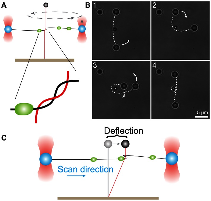
Figure 1. Detection of DNA-bound proteins using a scanning DNA loop and magnetic and optical tweezers.
(A) A DNA loop is created by moving the optically trapped beads (blue) around the magnetic-bead DNA tether (red). Zoom in shows the intertwined geometry of the DNA loop. The green dot depicts a bound protein. (B) Top view image series showing the formation of the DNA loop. The loop is made by rotating the beads trapped by optical tweezers around the magnetic bead which is located in the center of the image. The position of the DNA molecule is indicated by the white dashed line. The bead in the upper left corner functions as a reference and is stuck to the surface of the flow cell. (C) The loop is scanned in the horizontal direction by moving both optically trapped beads in concert. Upon encountering a bound protein the DNA cannot slide through the loop anymore and the magnetic bead tether is deflected.

