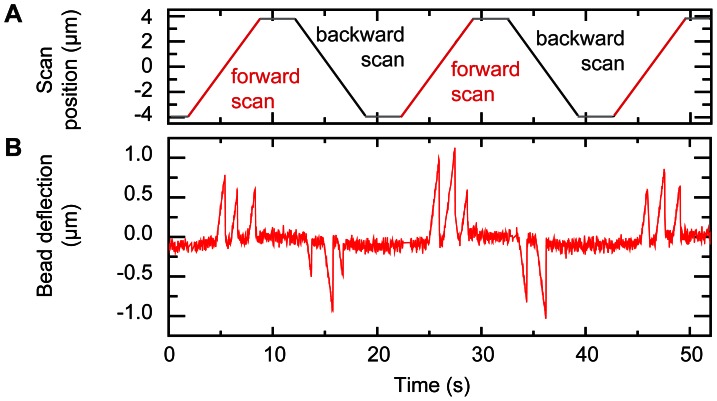
Figure 4. DNA-bound proteins are detected by scanning a DNA loop.
(A) Scan position defined as the mean position of the two optically trapped beads (red lines are forward scans, black lines are backward scans). (B) Magnetic bead deflection showing three spikes in both the forward and backward scans indicating the presence of DNA-bound proteins. The tension in both the magnetic bead tether and optical bead tether was set at 12 pN.

