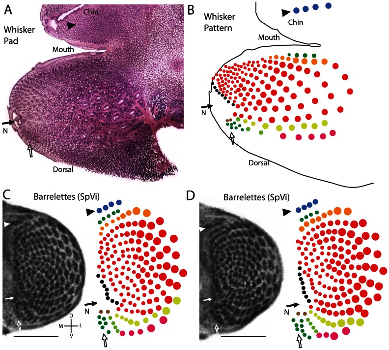
Figure 4. The proposed relationship between whiskers and barrelettes in the water shrew.
A. A single section from a juvenile water shrew whisker pad stained for hematoxylin and eosin shows the layout of the whiskers (sinus follicles) relative to mouth, chin, and nose. B. The distribution of whiskers as reconstructed from multiple sections. There were 4 whiskers located on the chin (blue circles). The nose (N), designated with a dark arrow, disrupted the pattern of whiskers providing a landmark reflected in the distribution of barrelettes at the medial extreme of SpVi. A small patch of whiskers was clustered dorsal to the nose (open arrow). C–D. Two examples of the barrelette pattern in SpVi from the left and right side of a single juvenile water shrew. The colored patterns of barrelettes to the right of the tissue sections were produced from examination of multiple sections. The proposed relationships between the landmarks noted on the whisker pad in A and B are indicated in C and D with corresponding colors, arrows, and arrowheads. Note that the whiskerpad illustrated in A and B is from a different case then the histology shown in C and D. Scale bars = 0.5 mm.

