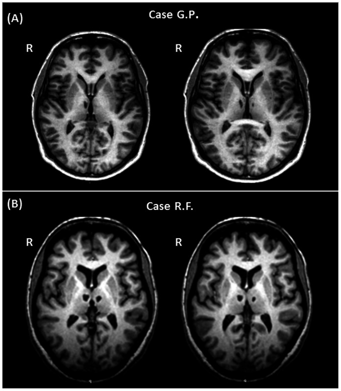
Figure 2. Macroscopic thalamic damage in the two studied patients.
Bilateral thalamic damage detectable on T1-weighted images of the two patients. G.P. shows a more asymmetrical involvement of the thalamus, with the right lesion considerably larger than the left one (A). Conversely, R.F. presents with a more symmetrical thalamic involvement, with both lesions showing a similar location and extension (B).

