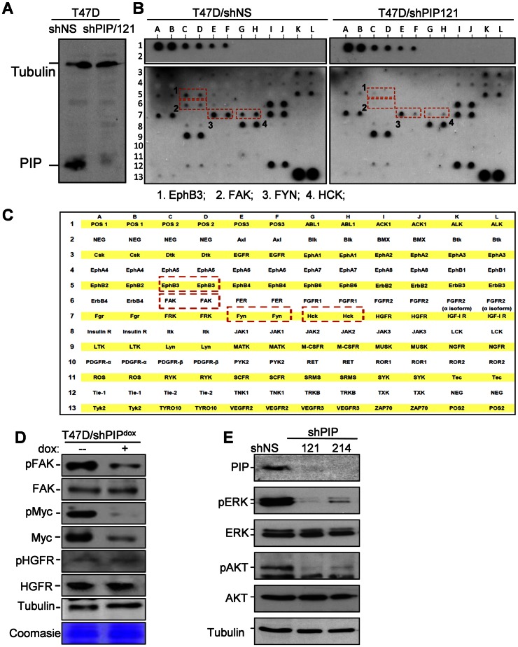
Figure 3. PIP silencing reduces phosphorylation of FAK, EphB3, FYN and HCK, and the downstream AKT and ERK1/2.
A, Immunoblot analysis of PIP in T47D cells expressing shRNA against PIP (shPIP/121) or a non-genomic target (shNS). B–C, Receptor tyrosine kinase (RTK) phosphorylation screen (B), and a map of the 71 RTKs and controls spotted on the antibody array (C). Cell extracts from T47D/shPIP/121 and T47D/shNS cells were hybridized to the membranes and RTKs showing reduced phosphorylation in PIP-silenced cells are indicated by red boxes. D, Western blot analysis of T47D/shPIPdox cells grown in serum-supplemented medium with dox or vehicle control. Antibodies were against [pTyr397]-FAK and pan FAK, [pThr58/pSer62]-Myc and pan Myc, as well as [Tyr1003]-HGFR and pan HGFR. Immunoblot of tubulin and a random coomasie blue-stained band are shown as loading controls. E, Western blot analysis of total and phosphorylated forms of AKT and ERK1/2 in cell lysates prepared from T47D cells expressing shRNA specific for either PIP (shPIP/121, shPIP/214) or a non-genomic target (shNS).

